What the Eye Anatomy Quiz measures
This quiz does not examine your eyes, certify you for clinical practice, or score your intelligence. It measures how much you agree with introductory eye anatomy literacy grouped four ways: cornea and front of eye about the clear window, sclera, iris, pupil, and first light entry; lens and focus about bending light, accommodation, and keeping lens location distinct from the cornea; retina, rods, and cones about photoreceptor placement, low-light versus color detail roles, and signal start toward the brain; and light path and study limits about cornea-to-retina tracing, diagram practice, and leaving diagnosis to professionals. You answer 22 statements from strongly disagree to strongly agree, with reverse-worded myths so agreeing with every shortcut cannot inflate every bar. Your answers also feed a second chart on closeness and trust habits, because health worry sometimes pushes people toward quick internet certainty. The total becomes an index from 0 to 100 where higher means more reported eye anatomy literacy on this screener, not proof you should skip care when symptoms scare you.
Cornea and front of eye
Clear cornea, sclera, iris, pupil, and how light first enters the eyeball.
Lens and focus
Lens placement, bending light, accommodation, and cornea versus lens roles.
Retina, rods, and cones
Back-of-eye layer, photoreceptors, fovea detail, and signal start toward the brain.
Light path and study limits
Cornea to retina tracing, retakes, labeled diagrams, and no DIY diagnosis.
How it works
Answer 22 agreement items
Rate each statement from Strongly disagree to Strongly agree. Tap Next after each answer.
Answers sort into four domains
Each answer feeds one of four eye anatomy groups, and every item also feeds a closeness and trust layer.
See your index and charts
You get a 0 to 100 index, a band, four domain bars, and a second chart for intimacy habits.
Take the Eye Anatomy Quiz
Answer 22 short statements to see your eye anatomy literacy index, four domain bars, and a closeness and trust chart.
What your result means
Your index is the average of all 22 answers, scaled from 0 to 100, with reverse-worded items flipped first. Higher means more reported eye anatomy literacy on this screener. Read light path and study limit bars together before you treat the total as clinical expertise.
| Band | Index range | Typical reading |
|---|---|---|
| Building eye anatomy literacy | 0 to 35 | Fewer agreed facts on cornea, lens, retina, light path, or study limits on this screener. |
| Mixed eye anatomy literacy | 36 to 65 | Solid facts in some domains with myths about lens location, rods and cones, or quiz-based diagnosis. |
| Strong eye anatomy literacy | 66 to 100 | Frequent agreement with accurate eye path trivia and healthy study limits. |
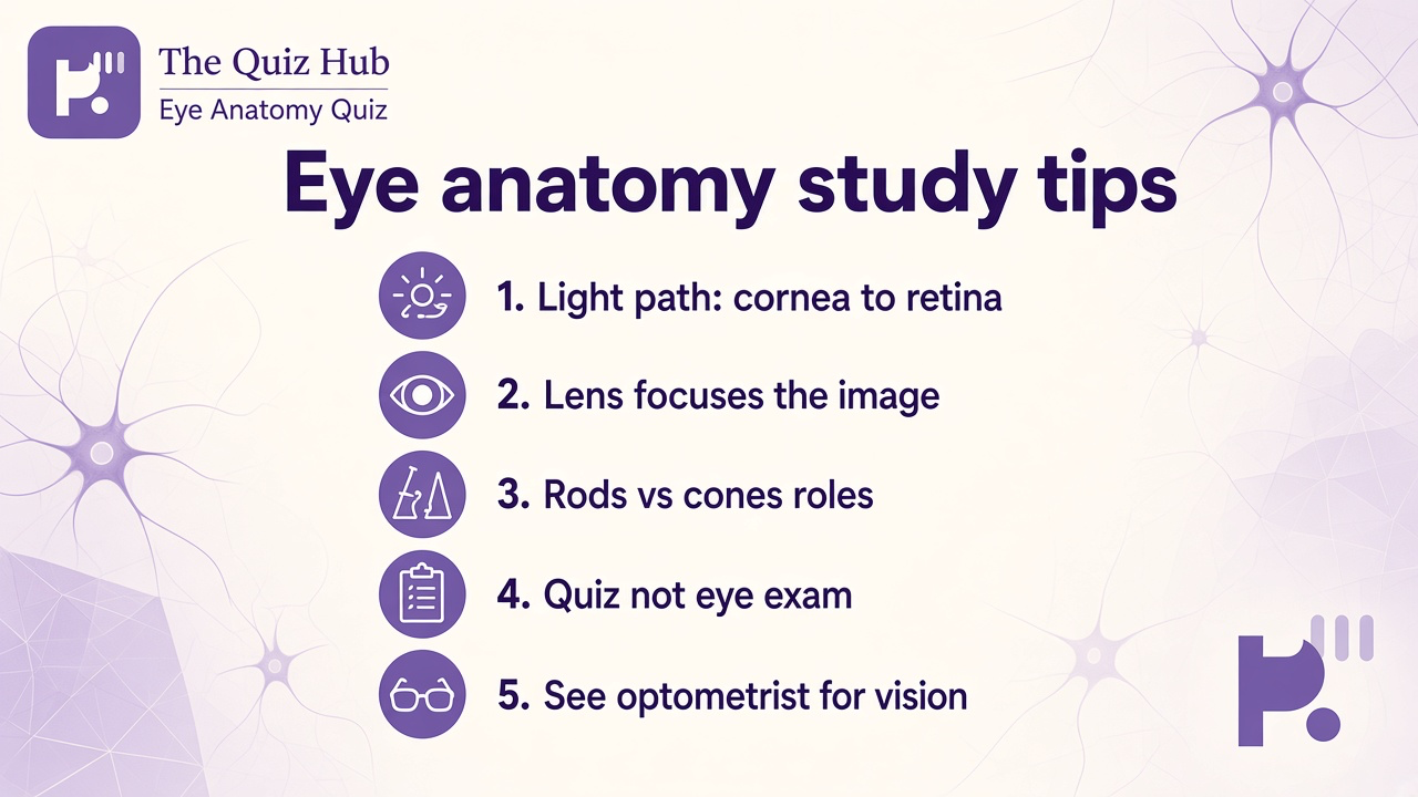
Tips at a glance
A quick takeaway graphic with topic tips (not your personal quiz scores). Start the quiz for your index, bands, and charts.
What this quiz can and cannot tell you
Can tell you
- Clarify cornea, lens, and retina names in plain language
- Highlight common cornea-lens and rods-cones myths worth unlearning
- Encourage diagram study and retake habits without shame
- Spark better questions during school vision or anatomy units
Cannot tell you
- Diagnose glaucoma, infection, or refractive error from trivia answers
- Replace dilated exams, imaging, or hands-on clinical testing
- Certify you for procedures, prescribing, or medical licensing
- Prove your eyes are healthy when blur, pain, or sudden loss appears
Eye anatomy literacy beyond one labeled poster
Students search eye anatomy when a biology unit hands them a cross-section and every region blurs together. The cornea is the clear front window that starts bending light; the sclera is the protective white you see around it. The iris colors the ring around the pupil, and the pupil size changes how much light enters. Mixing cornea and lens into one label is a trap quizzes flag on purpose because teachers want you to trace depth, not memorize shiny words.
The lens sits behind the iris and fine-tunes focus. Accommodation is the everyday term for changing lens shape to read a phone then look at a board. Camera analogies help as study shorthand even though living tissue is messier. Agreeing that only the cornea bends light or that the lens is optional decoration should not inflate literacy bars meant to track real structure habits.
The retina lines the back of the eye like film in a camera metaphor, except the biology is electrochemical. Rods support dim and peripheral vision; cones support color and sharp detail, especially in the fovea where cone density peaks. Signals move from photoreceptors toward the optic nerve and brain regions that build what you experience as sight. Claiming color happens only in the cornea or that rods and cones are optional is another myth pair worth rejecting before you retake.
Light path is the spine of intro units: cornea, pupil, lens, retina, then nerve traffic toward the brain. That order is simpler than every clinical nuance, but it beats memorizing names without direction. Study limits are the fourth domain because trivia scores tempt overconfidence. Retakes, flashcards, and labeled diagrams are normal class prep; they do not mean you finished optometry school. Agreeing that a perfect quiz replaces a dilated exam when vision blurs suddenly is another myth worth rejecting.
Use this screener as a warm-up, not a verdict. Pair it with our ear anatomy quiz when you want another sensory organ literacy break, the human body quiz for broader organ trivia, or the anatomy quiz when you want general structure recall. Browse the IQ and cognitive test hub for more low-stakes study games. Retake after you can trace light from cornea to retina without peeking; literacy grows when layers stay in order, not when you memorize that retina starts with r and hope the lens chapter never asks where rods live.
| Test | Focus |
|---|---|
| Ear anatomy quiz | Hearing pathways and pinna trivia, not cornea, lens, or retina photoreceptors |
| Human body quiz | Broad organ systems trivia rather than detailed eye light path literacy |
| Anatomy quiz | General anatomy recall rather than rod, cone, and accommodation depth |
Eye Anatomy Quiz FAQ
What does the Eye Anatomy Quiz measure?
It measures how much you agree with introductory eye anatomy literacy across cornea and front-of-eye basics, lens and focus, retina photoreceptors, and light path study limits. You get an index from 0 to 100 and four bars. It reflects self-reported knowledge on this screener, not a clinical skills exam or IQ score.
Is this quiz enough to diagnose vision problems?
No. It is study trivia and care literacy. Sudden blur, severe pain, flashes with curtain vision, or injury need an optometrist or ophthalmologist, not a high score here.
Why are some statements worded as myths?
Reverse-worded myths catch overconfidence. Agreeing that cornea and lens are the same structure, that the lens does not bend light, that rods and cones are optional, that color vision happens only in the cornea, or that trivia replaces dilated exams should not inflate your literacy index.
Does this cover every eye disease or surgical detail?
This screener stays at shared intro anatomy: regions, basic light path, rod and cone roles, and study habits. Advanced pathology, laser surgery, and full optometry boards belong in dedicated courses.
Who reviewed this quiz?
Dr. James Whitfield, PhD reviewed the educational framing, anatomy literacy limits, and result explanations. See our editorial policy for review standards.
Reviewed by